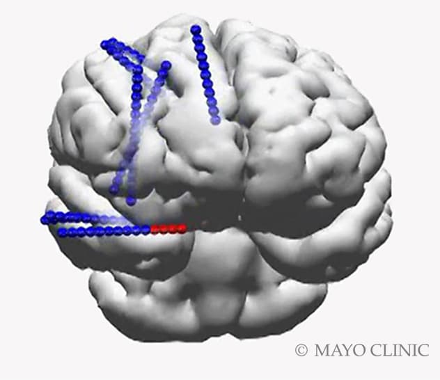


It may be noted that the higher frequency components were noted even when the scalp EEG did not show significant epileptiform activity. Conclusions: The components of the epileptiform activity are widely distributed in space, temporal and frequency domains, but specific components may be contributing to the significant pathophysiological processes. In the frequency domain, the intracranial EEG demonstrated a combination of high frequency (20-40 Hz and 50-150hz) and lower frequency (1-3 hz) components, with the higher frequency components showing lower power. Also, the epileptiform patterns tended to last longer over the grey mater regions. The spatial analysis showed that the intracranial EEG shows higher frequency patterns more prominently in the electrodes placed over grey mater regions of the brain compared to that over the white mater. Department of Neurology, Epilepsy Center, University Hospitals of Cleveland Medical Center, Cleveland, OH. The epileptiform activity (n= 20 identified from each patient) including sharp waves and spikes were analyzed for a period of around 2 seconds (+/-1 second) around the peak amplitude, for the frequency and time domain components. Results: The scalp EEG with sEEG (simultaneous recording) captured epileptiform patterns with similar abundance compared to initial scalp-only recording, suggesting that intracranial placement of the electrodes did not significantly affect the epileptiform activity. Stereo-EEG in Tuberous Sclerosis Complex. Further analysis of the signal was done in Matlab in temporal and frequency domains. The data was initially recorded in Nihon Kohden and exported in the ‘.eeg’ format and was imported to Matlab (Mathworks) for analysis, using the Brainstorm toolbox. The stereo EEG electrodes were placed on specific locations based on the hypothesis developed based on scalp EEG and brain imaging. These patients also had undergone initial EEG evaluation with scalp EEG. We use the stereo-EEG(sEEG) and scalp EEG recorded from widespread brain regions in identifying these relationships. Methods: We recorded the scalp (a modified 10-20 montage) and intracranial EEG (Stereo EEG), from approximately 140 electrode sites (implanted at multiple location) simultaneously, in patients undergoing stereo EEG monitoring as part of the presurgical work up for localization of epileptogenic zones for intractable epilepsy. We aim to establish the relationships between epileptiform activity recorded in intracranial and scalp EEG, both in frequency and time domains. Multiple approaches are used in determining the epileptogenic zone. Rationale: One of the important methods of treatment of intractable epilepsy is to remove the epileptogenic zones after identifying that location in brain.


 0 kommentar(er)
